WinCELL is a system for wood cell analysis
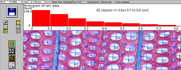
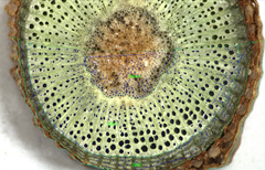
Image Analysis for Plant Science
img0
Regent Instruments Inc. since 1991


WinCELL


For sales informations,
contact us by email

sales@regentinstruments.com
cell analysis, wood cell, WinCELL, XLCell  
About WinCELL

WinCELL is an image analysis software program specifically designed for wood cells analysis. It can quantify the changes in wood structure over annual rings.

  cell analysis, wood cell, WinCELL, XLCell  

Anatomical wood cell analysis is an alternative to wood density analysis with x-rays (as done with WinDENDRO). Wood density, color, mechanical and chemical properties are in effect related to wood structure which in turns is related to climate. By measuring the radial cells (tracheids) size, distribution and their proportion to walls, wood quality can be assessed.

WinCELL measures wood cell morphology on thin wood slices cut with a microtome or, for larger cells like earlywood vessels of deciduous species, directly on wood surfaces*. Wood cells morphological data can be measured per annual ring.

Image analysers not made specifically for wood cells measurement are not usually able to produce data suited for dendrochronology studies. These systems lack some knowledge about annual tree rings formation and the structure of their cells (to compute the ring width for example). WinCELL has this kind of know-how built-in. It knows, for example, that a wall between two adjacent cells must be split in two to compute the cells length and earlywood or ring width. Its versatile settings allow to analyse different wood species (vessels of deciduous and radial row of conifer tracheids). It supports different automatic and interactive analysis modes. The latter allows you to select rows of cells to analyse them in a way that mimics the traditional trachedoid** method. WinCELL handles incomplete cells, those truncated by image boundary, so that they have no effects on the average cell measurements.

*With proper image acquisition device and sample preparation.
**Tracheidograms are curves of radial cell size variations in function of position in an annual ring.

Image Acquisition

Recent developments in high-quality affordable digital cameras have made anatomical wood cell analysis more accessible than ever.

Wood cell analysis is traditionally done on thin wood slices cut with a microtome. To acquire images of such samples, a microscope with a receptacle tube for a camera attachment and a camera adapter are required. Staining is sometime used to enhance lumen-wall contrasts when acquiring images in translucent light. This is the ideal setup to acquire very high resolution and precise images but the field of view is usually limited so that it is difficult or impossible to view entire rings.

cell analysis, wood cell, WinCELL, XLCell

Alternative sample's preparation methods and imaging over larger areas also exists. These are typically based on a scanner or a camera and macro lens and proper lighting. The difficulties with this approach are: to acquire images with good enough contrasts between lumen and walls, to do so without damaging them and to get enough resolution to accurately identify and analyse them. Their advantage is to allow to acquire images of a few annual rings per image, making their analyses easier.

cell analysis, wood cell, WinCELL, XLCell

WinCELL can analyse images acquired with scanners or digital cameras. The camera typically have a standard C-mount thread which allows it to be installed on a microscope using an adapter (C-Mount adapte). It can also be placed directly above the wood sample to acquire images with optional lenses. It is interfaced to computer via a fast USB connection. If you plan to use a scanner please inquire about the conditions before purchasing. WinCELL is TWAIN compatible, meaning it can acquire images from a few camera or scanner models.